(239)390-3339

Retina And Macula Information and Medical Treatment
Home
About Us
Contact
FAQ's
Seminar
Urgent Retina Disorders
Retinal Detachment symptoms and treatment

Retinal Detachment

Retinal Detachment in Bonita Springs, FL
Macula Disorders
  Serous Detachment of Retina
  Retinal Inflammation
  Retinal Infections
  Retinal Trauma
  Retinal Detachment
  Rhegmatogenous Retinal Detach...
  Retinal Tear
  Tractional Retinal Detachment
  Retinal Hole
  Retinal Lattice Degeneration
  Retinal Schisis
  Central Retinal Artery Occlusion
Vascular Retina Disorders
  Retinal Ischemia
  Retinal Disease & Pregnancy
  Retinal Hypertension
  Retinal Vasculopathy
  Retinal Macroaneurysm
  Retinal Microaneurysm
  Leukemic Retinopathy
  Diabetic Retinopathy
  Non Proliferative Retinopathy
  Proliferative Retinopathy
  Central Retinal Vein Occlusion
  Branch Retinal Vein Occlusion
  Branch Retinal Artery Occlusion
Chronic Retina Disorders
  Retinal Atrophy
  Retinal Dystrophy
  Retinal Toxicity
  Retinal Syphilis & Tuberculosis
  Radiation Retinopathy
  Retinal Toxoplasmosis
  Acute Retinal Necrosis Syndrome
  Birdshot Retinopathy
  Retinal Degeneration
Retina Procedures and Testing
  Retinal Laser Surgery
  Fluorescein Angiography
  3D Retinal Tomography
  Retinal Birefringence Scanner
  Retinal Treatments
Retinal Detachment treatment in Bonita Springs FL

Retinal Detachment

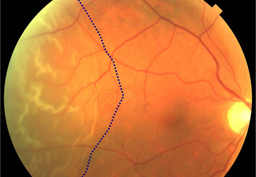
A Retinal Detachment is a separation of the retina from the underlying layer, called the retinal pigment epithelium. There are 3 main types: Serous Retinal Detachment, Rhegmatogenous Retinal Detachment and Tractional Retinal Detachment. The information you may have read or heard about a retinal detachment may have alarmed you. To know there is a chance of losing vision is frightening for anyone. Retinal detachment’s rarely result in a total loss of vision in the affected eye. They are most often successfully repaired with minimal vision loss. In years past, retinal detachment was thought by many to require immediate surgery or you would go blind. It is important to determine if your macula is impacted by the detachment. Commonly referred to as mac on or mac off detachments, the status of the macula prior to repair is the primary factor in determining your prognosis. While it is important to be evaluated as soon as possible after symptoms occur, it is not true that you will require surgery within 24 hours or you will go blind. One thing is true to all types of retinal detachments: you will need surgery to repair it.

Cause

 A retinal detachment can be caused by trauma, systemic illnesses, previous eye surgery, genetic tendencies or the aging process. The following are known factors that put you at risk for a retinal detachment:

Retinal tear – a tear is found in about 15% of all posterior vitreous detachments. A vitreous detachment is a normal part of aging. Most of the time it does not result in a retinal break or tear. If you have a tear, it is important to treat it with laser, since half of all untreated tears become retinal detachments.

Near sighted – particularly greater than -3 diopters. This makes you 10 times more likely to have a retinal detachment at some point in your life.

Lattice degeneration – is a preexisting condition in about 1/3 of retinal detachments. Lattice degeneration is areas of thinning around the peripheral retina and may have a lacy appearance. However, having laser prophylaxis around areas of lattice reduces your risk.

Cataract surgery – while cataract surgery increases your risk of having a retinal detachment, most patients that get a retinal detachment after cataract surgery had other preexisting risk factors.

Family history – If you have a parent or sibling who is known to be very near sighted, has lattice degeneration or retinoschesis, this multiplies your risk of retinal detachment.

Trauma – Any trauma to the eye may increase your risk of retinal detachment. If your wound penetrated the eye and caused bleeding, your risk increases over 70% for retinal detachment.

Systemic disease – common disorders such as diabetes and vascular disorders may increase your risk of retinal detachment, especially if they are not well managed and under control. There are also many rare disorders that have retinal detachment as an ocular side effect.

Rhegmatogenous retinal detachments are the most common type. These detachments are caused by a tear or break in the retina. Fluid can enter the opening and push the retina off its base, which is called the retinal pigment epithelium. These detachments respond well to surgical intervention.

Tractional retinal detachments are the next most common type. They are almost exclusively caused by systemic disease or a penetrating trauma to the eye. This type of detachment may include profound visual impairment.

Exudative retinal detachments are uncommon. They are caused by a breakdown of cellular structure or pressure gradient. This kind of retinal detachment is usually a result of a rare congenital or systemic disorder.

Symptoms

You may notice you do not have as much peripheral vision in the affected eye.  You may notice you have a dark area on the top, the bottom, or on a side of your vision. This is commonly described as a veil or curtain moving across your vision. Many patients have experienced an episode of flashing lights, or new floaters (dark spots) just before the retinal detachment happened.  If the detachment is at the top of your eye, this is called a superior detachment, however it will cause your vision loss to be in the lower portion of your visual field. When the lower portion of the eye is detached, this inferior defect will cause visual loss in the upper part of your visual field.

The severity level of your symptoms is related to the portion of the retina that is detached. For example, if there is a small area of retina detached at the edge, you would only see a tiny area of peripheral vision impacted, or nothing at all. This would be a macula on detachment, meaning the macula has been spared so far. You would need to cover your good eye to even notice it. Conversely, if the macula or entire retina becomes detached, you will notice a significant loss of vision in the affected eye. This is a macula off detachment. Even if a retinal detachment is small and unnoticeable at the onset, the area of detachment may enlarge if more fluid seeps underneath the retina.

Diagnosis

A retinal detachment is diagnosed during a dilated eye examination.  An optometrist, ophthalmologist, or retina surgeon may find the retina detachment, but only a retina surgeon can repair the damage and restore your vision. 
A retinal detachment is usually easily identified unless it is very small. Dr. Patel will do an examination called scleral depression to locate your tear or break. A scleral depression examination allows a view of the farthest edges of the retina, where tears are most commonly found. This exam is performed in a dark room, is mildly uncomfortable, but takes just a few minutes. A small instrument is used to apply pressure around the outside of your eye, while the doctor uses a special head set and lens to view the inside of your eye.  It is possible your tear will need to have laser treatment in the office on the day of your visit. This helps to secure the retina and prevent the detachment from spreading.
A color photograph of the inside of your eye may be taken to document the retinal detachment. A visual field test may be performed to assess the impact to your visual field.

In rare cases, a patient may present with significant vision loss because their retina has torn, and bleeding occurred from the opening. When blood mixes with the vitreous gel inside of the eye, you cannot see out of the eye, and the doctor cannot see inside the eye. In these instances, you would receive an ultrasound of the eye called a B-scan. The technology uses sound waves to image the back of the eye. This is a painless exam that allows the doctor to see if there is a tear or detachment behind the blood in the eye.

Treatment

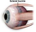
When a minimal retinal detachment is diagnosed very early, it may be possible to repair it with a laser treatment in our office. More advanced retinal detachments must be repaired with outpatient surgery. 

Several different surgical options exist for the repair of a retinal detachment. Your surgery will be tailored to address your specific needs. For instance, if you have already had cataract surgery in the past, a vitrectomy with scleral buckling may be the best option for you. If you have never had cataract surgery, or are a young adult at the time of your detachment, it might be preferable to place a scleral buckle without doing a vitrectomy.  A vitrectomy will cause a cataract to form postoperatively. If you have had cataract surgery, you cannot get another cataract.

A scleral buckle is a silicone band that is placed underneath the extraocular muscles, and sutured in place.


-


This band applies a tiny amount of tension to the middle of the eye, which in turn helps to hold the retina in place, according to the formula T=P/R (tension = pressure divided by radius of the eye). The band is not visible on the outside of the eye, and it stays in place permanently. 

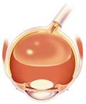
A common procedure included in any type of retinal detachment surgery is the placement of a gas bubble. The SF6 gas bubble is short acting, and will be in place for a few weeks. The C3F8 gas bubble is longer acting, and will be in place for 8-12 weeks. The length of time the bubble stays in the eye is dependent on the body’s ability to metabolize or absorb the bubble according to chemical formulas. The gas bubble is a critical part in keeping the retina in place during reattachment. These gases are lighter than water, and will rise to the top of a fluid filled container. Therefore, when you sit up straight and look forward, the gas bubble is at the top of your eye. The retina is on the back wall of the eye, so to make the bubble apply pressure to the retina, your face must be positioned parallel to the floor.


You will be required to hold this position for several days while you are awake. You will be able to take breaks for about 10 minutes every hour to eat and drink, etc. You will need to sleep on one side for the entire time you have the gas bubble. You may not sleep on your back. This is to prevent the gas bubble from pushing against the front of your eye, where it could cause damage.

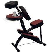
Special chairs are available for rent to help you maintain the proper positioning. The first week after surgery is the most restrictive. Your ability to maintain a face down position will impact the quality of vision you have postoperatively.


You will not be able to participate in strenuous activities for several weeks after surgery. You will be able to resume most of your normal routine after the first week. Driving will depend on your level of comfort, since the gas bubble will impair your vision in the affected eye.

While this is a difficult time for our patients, most are able to regain nearly the same level of vision they had prior to the retinal detachment. It is rare that anyone goes blind in the eye that suffers a detachment.

Prevention

 Some underlying eye conditions are known to have a higher incidence of retinal detachment.  In many cases, a laser treatment to weak or compromised areas of the peripheral retina may reduce the risk of retinal detachment.  Safety glasses and goggles help prevent trauma to the eye that can cause retinal detachment.  A healthy lifestyle and careful management of diabetes also helps to reduce your risks.

Contact Us

Please contact us at (239)390-3339 or click here to use our contact form
Retinal Detachment symptoms and treatment


Retinal Detachment in Bonita Springs, FL
  Macular Dystrophy
  Macular Serous Detachment
  Macular Edema
  Macular Degeneration
  Macular Ischemia
  Macular Hole
  Macular Epiretinal Membrane
  Macular Pucker
  Macular Traction
  Macular Telangiectasias
  Acute Macular Neuroretinopathy
  Macular Cystoid Macular Edema
  Macular Dragging
  Macular Coloboma
Retina and Macula
Retina and Macula Treatment Retina and Macula Privacy Retina and Macula Disclaimer


Macula and Retina

Maculas and RetinasMacula and Retina FLMacula and Retina Florida
Retina NaplesRetina Naples FLRetina Naples Florida
Retina Fort MyersRetina Fort Myers FLRetina Fort Myers Florida
Retina Bonita SpringsRetina Bonita Springs FLRetina Bonita Springs Florida
Retina CitiesRetina Cities FLRetina Cities Florida
Macula NaplesMacula Naples FLMacula Naples Florida
Macula Fort MyersMacula Fort Myers FLMacual Fort Myers Florida
Macula Bonita SpringsMacula Bonita Springs FLMacula Bonita Springs Florida
Macula CitiesMacula Cities FLMacula Cities Florida
Retina and Macula Naples Retina and Macula Fort MyersRetina and Macula Bonita Springs